
Complexion Analysers (Dermoscopes)
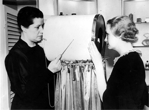
On September 6, 1939 the firm of Helena Rubinstein held a press conference to unveil her new complexion analyser, the ‘Polaroid Dermascope’, the first device that used polarised light to examine the skin. At the press conference was Dr. Martin Grabau, from the Polaroid Corporation and Mala Rubinstein, representing Helena Rubinstein, Inc.
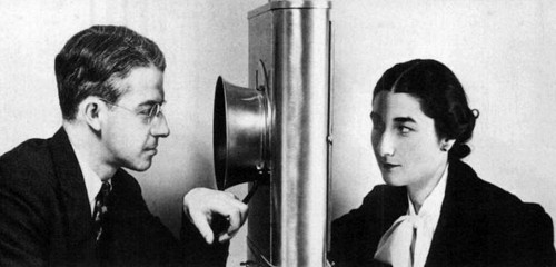
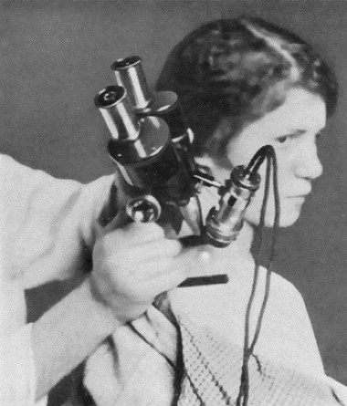
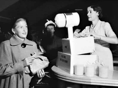
Above: 1939 The Polaroid Dermascope ‘skin analyser’ demonstrated by Polaroid’s Dr. Martin Grabau on the skin of Mala Rubinstein.
Although it looks nothing like current forms, the device had some functional similarities with modern dermatoscopes.
Dermatoscopes
The first dermatoscope was developed In 1920 by the German dermatologist Johann Saphier. The device, which he called a ‘dermatoskope’, was a rather cumbersome instrument consisting of a binocular microscope with an in-built light source. He used it to make detail observations of the skin. Dermatologists largely ignored Saphier’s ideas, relying on their eyes and the occasional use of a magnifying glass, rather than on such a specialist instrument.
In the 1950s, Leon Goldman of the United States wrote a series of papers describing how he used a similar instrument to investigate pigmented skin lesions and he develop a portable device in 1958 to help with his research. Goldman described his studies as ‘dermoscopy’ rather than ‘dermatoscopy’. This probably led to some dermatologists referring to dermatoscopes as dermascopes, although this occurred well after Helena Rubinstein’s use of the term. Others followed Goldman’s work but it would not be until the 1980s, when skin cancers became more common, that these portable devices would gain any real traction with dermatologists.
Dermatoscopes are mainly used to help distinguish between benign and malignant skin cancers, with a number of researchers suggesting that they increase the accuracy of identification. As well as magnifying the skin so that surface details are easier to see, they also make the subsurface of the skin more visible by reducing surface skin reflection. This can be achieved by using an immersion oil technique to create a direct contact with the skin and/or by viewing the skin under polarised light.
Light is either reflected, dispersed, or absorbed by the stratum corneum because of its refraction index and its optical density, which is different from air. Thus, deeper underlying structures cannot be adequately visualized. However, when various immersion liquids (immersion oil, alcohol, or ecography gel) are used, they render the skin surface translucent and reduce the reflection, so that underlying structures are readily visible. The application of a glass plate flattens the skin surface and provides an even surface. Optical magnification is used for examination. Taken together, these optical means allow the visualization of certain epidermal, dermo-epidermal, and dermal structures in contact dermoscopy.
Recently, newer hand-held dermatoscopes use a polarized light filter to significantly reduce irregular light reflection allowing underlying structures to be visualized without the need of applying an immersion liquid.(Roldán-MarIín, Puig & Malvehy, 2012, p. 150)
Like many modern dermatoscopes, Helena Rubinstein’s skin analyser also used polarised light to see sub-surface detail invisible to the naked eye, predating these modern instruments by many decades.
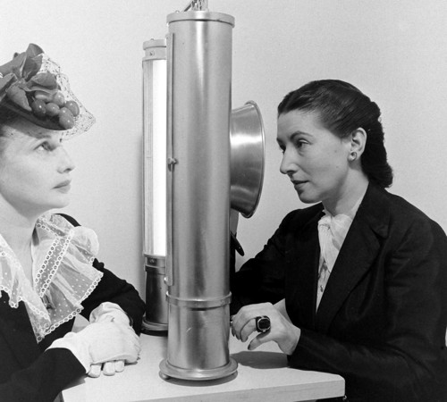
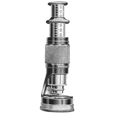
Above: 1941 Helena Rubinstein’s Polaroid Dermascope.
Given that polarised light was a rather novel idea in 1939 – polarised sunglasses having only just appeared – a detailed explanation was given to the press on how the instrument operated, using language they would hopefully understand.
By using the new instrument, called the “Polaroid Dermascope,” the makers state an observer can examine the lower layers of the skin almost as clearly as if the outer skin surface were not there. Then, by moving a control lever, he can throw the outer surface into even greater prominence than under ordinary light, for examination of surface defects.
Many skin conditions, partly or wholly invisible under ordinary light, it is stated, are clearly detected with the new instrument. The device is a development of the technical staff of Helena Rubinstein. It uses the same Polaroid light-control material applied in three-dimensional movies, antiglare glasses and desk lamps, and proposed for eliminating headlight glare. It is the first of a series of examination and inspection devices employing a similar principle, to be brought to use by the Polaroid Company. Others, now being developed, include instruments specially designed for use by dermatologists, opthamologists, dentists, and other medical specialists, criminal investigators and art experts.
The Polaroid Dermascope consists of two sets of light sources, one fitted on either side of the instrument, fitted with reflectors which concentrate the light on the face. Over those sources are place Polaroid filters. The raw light comes in one side of these transparent filters, like a bundle of round rods and comes put the other like a bundle of flat ribbons. The vibrations are shaped by the billions of invisible crystals embedded in every Polaroid sheet.
These ribbons of light fall upon the skin. According to the maker’s description, those that bounce from the top surface, as shine, retain their ribbon-like form. Those that penetrate the skin, lose their ribbon form and are converted into ordinary light.
By looking through the Polaroid observation plate, and turning this plate so that its “optical slots” are at right angles, or crosswise, to the ribbons that are bouncing from the surface as shiny reflection, the observer, it is pointed out, can block them off altogether. He sees only the light which has penetrated down into the lower layers of the skin, where the tissues have broken up the Polaroid light ribbons and converted them back into ordinary light. The operator sees the sub-surface layers clearly because the ribbons of light which make up the shiny top-surface mask are blocked by the crosswise optical slots of the Polaroid viewing plate.
The device is equipped with a lens to give the operator an enlarged view of the subject’s skin.(AP&EOR, 1939)

Left: Skin as seen with normal light. Right: Skin viewed through the Polaroid Dermascope.
Despite the fact that the device was able to see skin features not readily apparent to the naked eye it seems unlikely that the Polaroid Dermascope enabled an experienced beauty therapist to learn a lot more about a client’s skin that she could see with her naked eye. The machines operated more as a novelty when displayed in department stores, helping to convince clients that a recommended treatment regime was scientifically valid, a situation strengthened when the demonstrators were men. As far as I can tell they were not used in salons. Helena Rubinstein must have found these devices useful in generating sales as this was not her first use of an optical skin analyser. That came some 5 years earlier with the ‘Derma-Lens’.
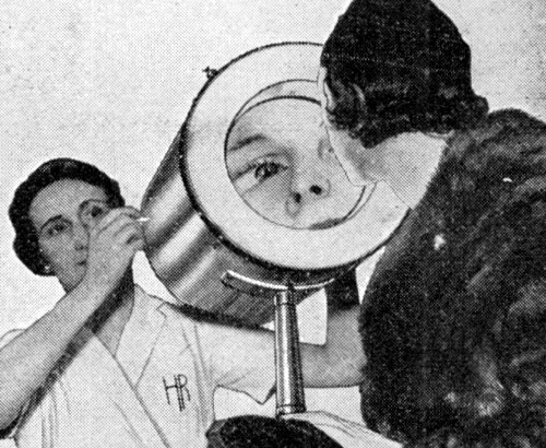
Derma-Lens
The first model of the of the Derma-Lens was a rather cumbersome apparatus. it used lights and curved mirrors to enable the therapist and the client to see a magnified image of the client’s face at the same time.
1934 Helena Rubinstein Derma-Lens.
A new device for scientific analysis of complexions, known as the “Derma-Lens,” has been designed by Helena Rubinstein, Inc. New York, and is being used in various department stores by representatives of the company to determine the exact beauty treatment required by women. … Moveable light inside the “Derma-Lens” make it possible to focus attention on special skin areas.
The apparatus is said to gauge the exact texture and condition of the skin, and enables the operator to peer critically into the skin where flaws develop and advise women accurately as to the exact corrective home beauty care the individual complexion requires. It is claimed that the “Derma-Lens” reveals the true flesh tone of the skin and thus provides the basis for the selection of perfectly harmonizing cosmetics.(AP&EOR, 1934)
Later models were more elegant, smaller and cheaper and operated like a more complicated version of a modern salon magnifying lamp, the main difference being that the client was also able to see the magnified image.
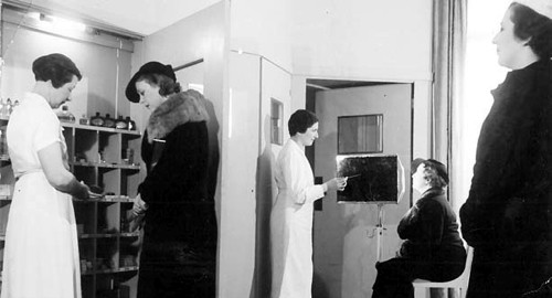
Derma-Lens as used in Helena Rubinstein’s Insitut de Beauté, Paris.
1935 Later model Derma-Lens.
1936 This model is referred to as a Dermascope when reported in Modern Mechanix even though it is clearly being staffed by a therapist from Helena Rubinstein. The client was also able to see her face magnified in a mirror that was well lit by a circular fluorescent light. The caption reads: “This device brought out in England especially for use in beauty parlors, enables a client to examine face, hair, etc., under high magnifying power, which will bring up blemishes in a way to sell services and cosmetics. It evidently contains a concave mirror and special illuminating means”.
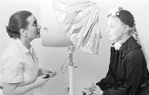
1937 Derma-Lens as used in Helena Rubinstein’s New York salon. This model is similar to the one above with a large, circular, fluorescent light source at the back that illuminates every facial blemish. The client’s head would normally be covered with the drape.
Other devices
Helena Rubinstein was not the only cosmetic firm to use optical devices to analyse the skin’s complexion. Images of scientists peering at skin through ‘scopes’ appear occasionally in cosmetic advertising and I have included a few examples for examination. In the 1950s, other cosmetic companies also used complexion analysers in department store demonstrations to help generate sales but the practice seems to have died out after that.
Salon usage
Optical complexion analysers that function as a dermatoscope were not widely used in salons, partly because of their size and expense but also, as mentioned earlier, because they did not tell an experienced therapist much more than they could see with their naked eyes – the same reason they were not rapidly adopted by dermatologists. As a selling device most of the less-expensive forms also suffered from a major limitation – unlike Rubinstein’s Derma-Lens – namely that the client could not see the skin problems the operator was viewing and describing. This limitation has been overcome in recent years with the arrival of computers and digital photography. The development of small, relatively cheap dermatoscopes that can be attached to a smartphone or tablet, allows magnified images to be taken with the built-in camera and then shown to the client. This opens up more possibilities for them being used in salon consultations.
First Posted: 14th January 2014
Last Update: 16th March 2023
Sources
The American perfumer & essential oil review. (1934, 1939). New York: Robbins Perfumer Co.
Earls, A. R., & Rohani, N. (2005). Images of America. Polaroid. Charleston, SC: Arcadia Publishing.
Lee, J. B., & Hirokawa, D. (2010). Dermatoscopy: An overview—Part I: Nonmelanocytic lesions. Skinmed. 8(5), 265-272.
Roldán-MarIín, R., Puig, S., & Malvehy, J. (2012). Dermoscopic criteria and melanocytic lesions. Giornale Italiano di Dermatologia e Venereologia. 147(2), 149-59
Saphier J. (1921). Die dermatoskopie. IV. Mitteilung. Archiv für Dermatologie und Syphilis. 136(2), 149-58.
Skin-tone analyzer selects right color for face cosmetics. (1939). Popular Science. Retrieved January 2, 2014, from http://blog.modernmechanix.com/skin-tone-analyzer-selects-right-color-for-face-cosmetics/

Edwin Land [1909-1991]. The inventor of the sheet polarizer as used in Polaroid Sunglasses and the Helena Rubinstein Polaroid Dermascope. He later developed the Land (Polaroid) Camera.
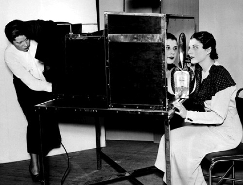
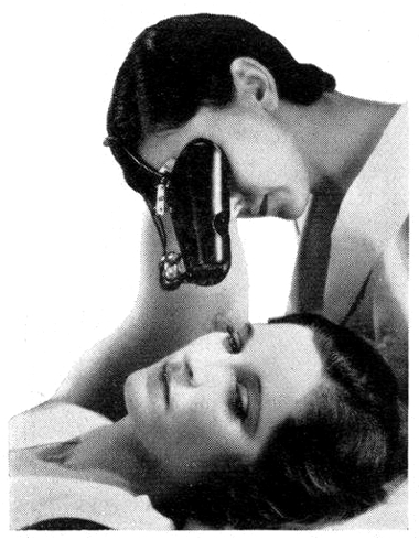
Above: 1939 Dr. Grabau standing behind the Polaroid Dermascope (Earls & Rohani, 2005) showing the client side of the device.
1921 A modified binocular microscope (dermatoscope) as used by the German dermatologist, Johann Saphier (Saphier, 1921).
1922 Johann Sapier’s design for a less cumbersome dermascope.
1931 Ambrosia Cream with an image of a scientist using what appears to be a type of dermatoscope to examine the skin.
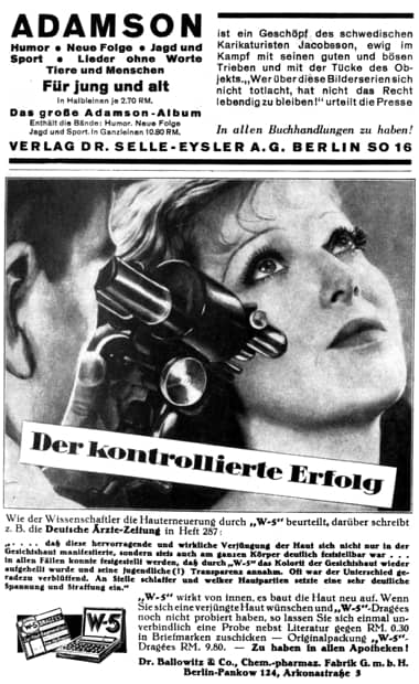
1933 Dr. Ballowitz & Co. W-5 Dragees.
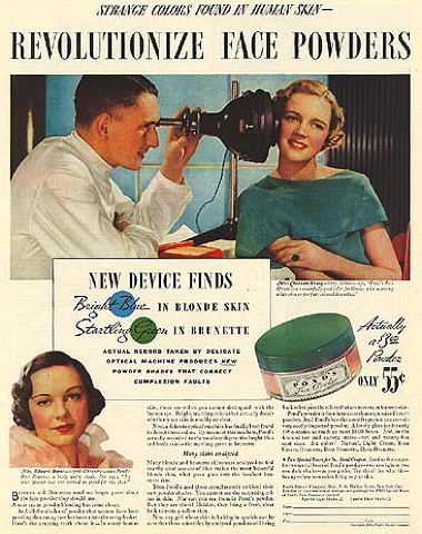
1934 Pond’s Color-Scope scientific shade detector.
1934 Marinello Dermascope.
1936 Marinello Dermascope as used in the Marinello salon at 36, Avenue Hoche, Paris. Some Marinello salons had been using these devices from 1931.
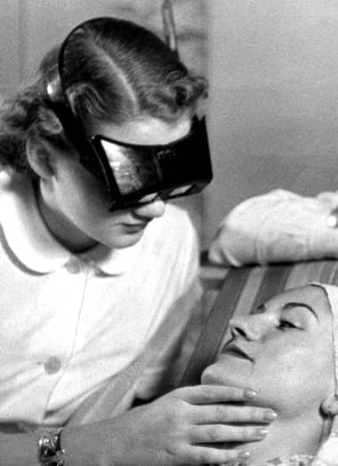
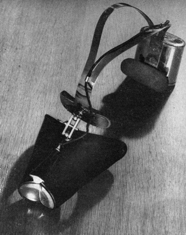
1937 Magnifying goggles being used in a German beauty salon.
1939 Skin-Tone Analyzer. “To determine the exact cosmetic color that will best blend with individual skin tones, a novel analyzing apparatus has just been introduced for beauty-shop use. A complicated system of lenses, prisms, and a polarizing screen is used to throw a tiny rainbow of light onto the skin of a customer. Analysis of the basic color tone of the skin is then made by noting which shade of the spectrum is most absorbed and which is most reflected” (Popular Science, 1939).
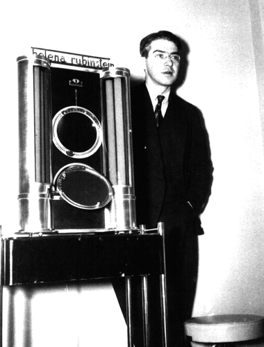
1940 Helena Rubinstein’s Polaroid Dermascope.
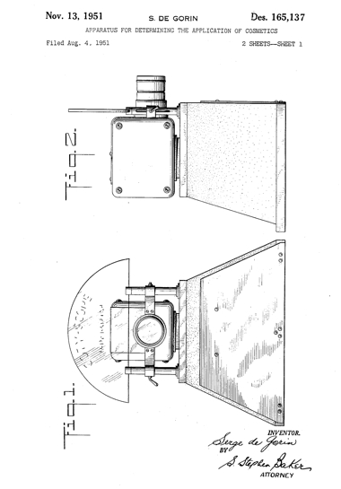
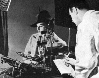
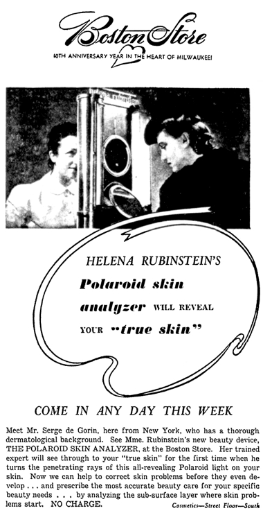
1951 Serge de Gorin patent for the Coty-Scope. Described as a physicist or dermatologist in Helena Rubinstein advertisements, Serge de Gorin demonstrated her Polaroid Dermascope in the 1940s in American department stores. He did similar work for Coty in the 1950s presumably using this device he patented.
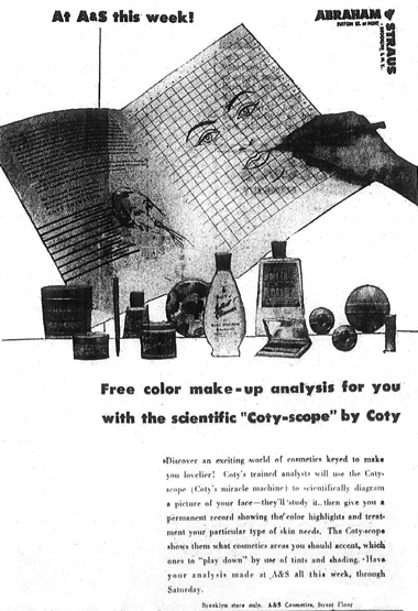
1953 The Coty-Scope.
1953 A Skin Color-O-Graph machine used by the Paul Duval company in a department store demonstration in Australia.